| Email:Mannan.Ali@physics.org | Chapter 2: Magneto Optical Kerr Effect (MOKE) |
|---|---|
| Web: http://members.xoom.com/MannansZone/thesis.html |
2.0 Magneto Optic Kerr Effect (MOKE)
2.1 Introduction
Magneto Optical effects in magnetic materials arise due to the optical anisotropy of the materials. The source of this optical anisotropy is the magnetisation M within surface domains which can be influenced by external forces such as magnetic fields. The optical anisotropy alters the state of linearly polarised light which is reflected off magnetic materials. These effects are generally known as the Magneto-Optical Kerr Effect (MOKE) which were observed by John Kerr in 1887, and are analogous to the Faraday effect where the polarisation of the light is rotated through a transparent material subjected to a magnetic field as observed by Michael Faraday in 1845.
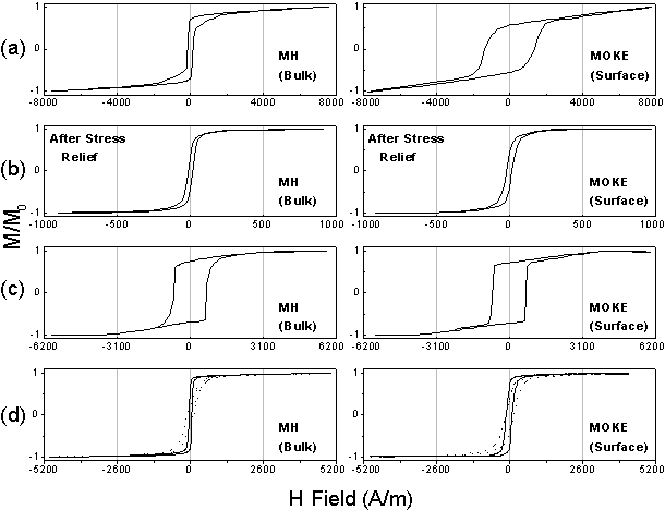
The magneto optical effects are characterised by the Kerr effect being proportional to the magnetisation. This makes it particularly useful in the study of surface magnetism since it is highly sensitive to the magnetisation within the skin depth region, typically 10-20nm in most metals [Bland et al (1989)]. The effect has been utilised to obtain hysteresis loops or domain images and is a relatively simple technique to implement. It has the ability to probe the magnetisation in very small regions of a material, such as wires or patterns [Shearwood et al (1996)], or in real device applications [Karl et al (1999)]. MOKE has emerged as an important technique in the study of surface magnetism. It has been used extensively to characterise magnetic materials, especially in the field of magnetic thin films. The Kerr effect is also the basis of the commercially available magneto optical drives.
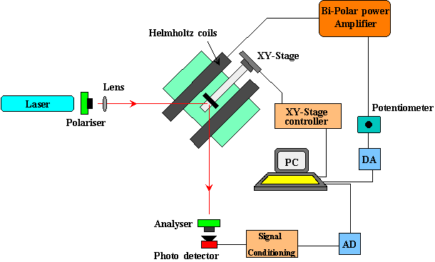
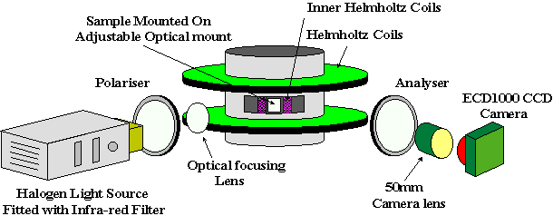
Part of this study has been involved with the construction of a MOKE magnetometer and MOKE imaging system, in order that the amorphous ferromagnetic thin films could be studied. The MOKE magnetometer provided information in the form of hysteresis loops, whereas the imaging system provided magnetic information by means of domain images. The two systems were built to overcome the limitations of the Inductive Magnetometer which was designed primarily for studying amorphous, ribbon based materials. The principles of MOKE are discussed here only qualitatively; this account is not intended to be complete and rigorous, but to provide an overview for the reader.
| Growth and study of magnetostrictive FeSiBC thin films, for device applications, Mannan Ali (1999) | 3 | |
|---|---|---|
| (Online Copy) |
| Email:Mannan.Ali@physics.org | Chapter 2: Magneto Optical Kerr Effect (MOKE) |
|---|---|
| Web: http://members.xoom.com/MannansZone/thesis.html |
2.2 Principles of MOKE
Magneto Optical Kerr effects are generally described macroscopically by dielectric tensor theory [Zak et al (1990)], or the effects can also be described microscopically, where the coupling between the electric field of the light and the magnetisation occurs by the spin-orbit interaction [Daalderop et al (1988)]. In the present work the effects are described less formally, in a pictorial fashion, using the idea of a Lorentz force. To understand the magneto optical Kerr effects, one needs to understand the terminologies associated with the effect, how the state of polarisation of reflected light is dependent upon the initial polarisation and the magneto optical geometry in which it is being used.
Light is a transverse electromagnetic wave which can be manipulated optically into plane, circularly or elliptically polarised light (Fig. 2.1). Generally, the plane of polarisation is the plane which contains the electric field E and the direction of propagation. However in some texts [Corson & Lorrain (1970)], the definition of plane of polarisation refers to the plane containing the B field. Any reference to the plane of polarisation in the present work will assume the former definition. If the electric field is polarised in the plane of incidence, it is referred to as p-polarised light as shown in Figure 2.2. Conversely, if the electric field is polarised perpendicular to the plane of incidence, then it is referred to as s-polarised light. The plane of incidence is also known as the scattering plane - the plane which contains the incident and reflected light beam. Circularly polarised light can be further referred to as L-circularly polarised and R-circularly polarised light, where L and R signify the electric field rotating in either a clockwise or an anticlockwise direction with respect to the direction of propagation.
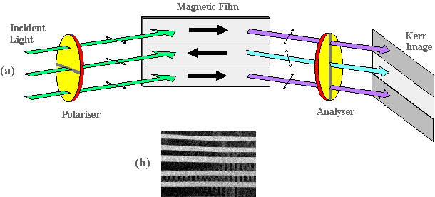
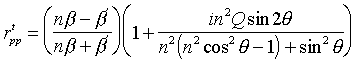
Plane polarised light which is reflected off a metallic surface, is generally elliptically polarised. However if the incident light is either p or s-polarised, then the reflected light will still be plane polarised upon reflection (p or s) [Hecht (1989)]. This is because the reflecting surface is a plane of symmetry for the system. This symmetry is destroyed in the situation where plane polarised light is reflected off a magnetised surface. When p-polarised light is reflected off a magnetic surface, the reflected light has a p-component as in the ordinary metallic reflection but, in addition, a small s-component also appears in the beam. In general, this second electric field component is out of phase with the reflected p-component. This causes the light to become elliptically polarised with its major axis rotated from its initial incident polarisation plane. This magneto optic interaction is shown schematically in Figure 2.3. A similar effect occurs for s-polarised light. The two effects are know as the Kerr ellipticity and the Kerr rotation. As mentioned earlier, the effects are described macroscopically using dielectric tensor theory. In this theory, plane polarised light is viewed as being
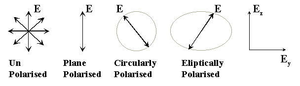
Figure 2.1: Polarisation of light.
| Growth and study of magnetostrictive FeSiBC thin films, for device applications, Mannan Ali (1999) | 4 | |
|---|---|---|
| (Online Copy) |
| Email:Mannan.Ali@physics.org | Chapter 2: Magneto Optical Kerr Effect (MOKE) |
|---|---|
| Web: http://members.xoom.com/MannansZone/thesis.html |
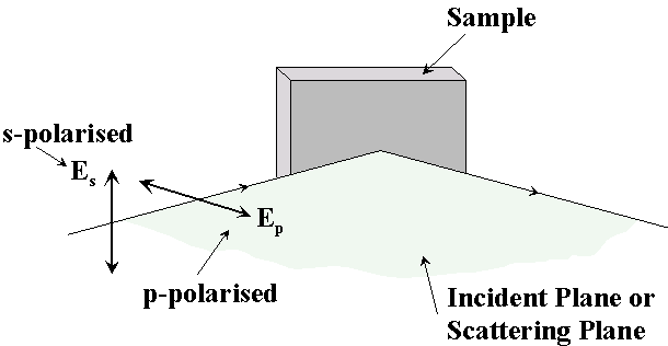
Figure 2.2: Illustration of p-polarised and s-polarised light.
made up of the superposition of two circular components, L and R-circularly polarised light. The magnetic medium has different refractive indices for these two polarised modes. Therefore the two circular modes travel with different velocities and attenuate differently in the material. Upon reflection from the material, the two modes recombine to produce the Kerr rotation and ellipticity. The macroscopic description of Kerr effects relies on the two modes having different refractive indices within the material. The general form of the dielectric tensor which represents the effects of a magnetic medium is given by [see Zak et al (1990) for details]
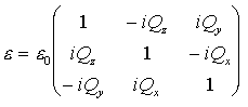 | (2.1) |
where Qx,y,z is the Voigt magneto optic constant which describes the magneto optical effect. This Voigt term is to the first order proportional to the magnetisation of the material. It is this complex Voigt term (the off diagonal terms) which generally modifies the polarisation. Basically, what MOKE measures directly, is the magneto optic response of the medium, which is a change in the incident polarisation of the light. This magneto optic response consists of two parts: a change in the polarisation of the in-phase component of the reflected light which gives rise to the rotation, and a change in the polarisation of the out-of-phase component of the reflected light which gives rise to the ellipticity.
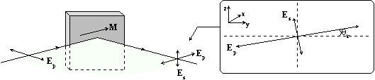
Figure 2.3: Reflection of p-polarised light of a magnetic sample.
| Growth and study of magnetostrictive FeSiBC thin films, for device applications, Mannan Ali (1999) | 5 | |
|---|---|---|
| (Online Copy) |
| Email:Mannan.Ali@physics.org | Chapter 2: Magneto Optical Kerr Effect (MOKE) |
|---|---|
| Web: http://members.xoom.com/MannansZone/thesis.html |
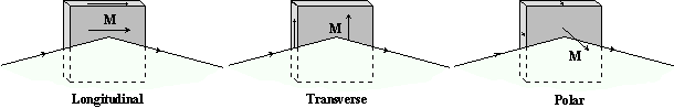
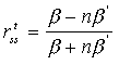
Figure 2.4: Longitudinal, transverse and polar Kerr effects.
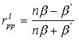
There are principally three Kerr effects which are classified depending upon the magneto optic geometry being employed. These are shown in Figure 2.4. The effects are dependent on the orientation of the magnetisation with respect to the incident and sample planes. In the longitudinal Kerr effect, the magnetisation is in the plane of the sample and parallel to the incident plane. In the transverse Kerr effect, the magnetisation is also in the plane of the sample, but is perpendicular to the incident plane. In the polar Kerr effect, the magnetisation is perpendicular to the sample plane and is parallel to the plane of incidence. It should be noted the Kerr effect will occur for any arbitrary direction of magnetisation within the sample. Consideration of these three magneto optic geometries simplifies the understanding of the Kerr effect. The longitudinal and transverse Kerr effects are generally used to study the in-plane magnetic anisotropy, whereas the polar configuration is used to study thin films, which exhibit perpendicular anisotropy. The thin films investigated in this study on the whole only exhibited an in-plane magnetic anisotropy and therefore the polar effect was not used. Upon refection the longitudinal and polar Kerr effects, generally, alter the polarisation of the incident light from plane to elliptically polarised with the major axis rotated (Kerr rotation). In the transverse effect there is no change in the polarisation of the incident light. This is more clearly illustrated in Figure 2.5, where a vector representation using the idea of a Lorentz force indicates how p and s-polarised light interact in the three magneto optic geometries. The electric field of the plane polarised light which is incident upon the material, can be thought of as exciting the electrons so that they oscillate parallel to the incident polarisation. This gives rise to the normal component (EN) of light in the reflected light. The additional Kerr component, EK, arises because of the Lorentz force. The Lorentz force induces a small component which is perpendicular to both the primary motion (normal component) and the direction of the magnetisation. Generally, the two components are not in-phase and it is the superposition of these two components which gives rise to a magnetisation dependent rotation of the polarisation. In the longitudinal and polar Kerr effects (Fig. 2.5a,b), p or s-polarised light will generally become elliptically polarised with its major axis rotated (Kerr rotation). This is a consequence of an orthogonal electric field component being induced because of the Lorentz force. The directions of the Lorentz force, and therefore the induced components, are shown by the dashed arrows (EK). The Kerr effect diminishes as the angle of incident approaches the normal to the sample plane in the longitudinal effect because either the Lorentz force vanishes (p-polarised) or points along the direction of the light (s-polarised). This is not the case for the polar Kerr effect because the magnetisation is out of the sample plane and a Lorentz force always exists at normal incidence. The polar effect is independent of the incident polarisation at normal incidence. The angle of incidence generally tends to be independent of the incident polarisation
| Growth and study of magnetostrictive FeSiBC thin films, for device applications, Mannan Ali (1999) | 6 | |
|---|---|---|
| (Online Copy) |
| Email:Mannan.Ali@physics.org | Chapter 2: Magneto Optical Kerr Effect (MOKE) |
|---|---|
| Web: http://members.xoom.com/MannansZone/thesis.html |
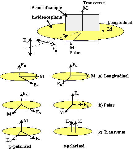
Figure 2.5: A schematic representation of the Magneto Optic interaction using the idea of a Lorentz force. The normal component (EN) of light is indicated by the solid lines, the Kerr component (EK) and the direction of the Lorentz force is indicated by the broken lines.
at normal incidence. The angle of incidence generally tends to vary in the range 5-600 depending on the experimental arrangement for the longitudinal and transverse modes. In most instances the angle of incidence is fixed by the constraints of the experimental layout. It has been shown experimentally [Deeter & Sarid (1988)] that the angle of incidence does have a small effect on the magnitude of the Kerr rotation. The polar Kerr effect is usually an order of magnitude larger than the longitudinal Kerr effect. The transverse effect involves no change in polarisation, since there is either no Lorentz force present (s-polarised) or the induced component (p-polarised) has the same polarisation as the incident polarisation (Fig. 2.5c). The transverse effect involves a change in the intensity of the light (Kerr reflectivity). The intensity changes are dependent upon the component of magnetisation perpendicular to the plane of incidence. There is no Kerr ellipticity since M´E induces a component which is in the plane of incidence. The induced Kerr component and the normal component give rise to a change in the amplitude. One only sees a Kerr rotation if a Lorentz force is present. In general either s or p-polarised light is used. This is because any change in the polarisation of the light will be a result of the magnetisation, since in an ideal situation there will be no change in the polarisation of light for either s or p-polarised light reflected off a non-magnetic surface.
| Growth and study of magnetostrictive FeSiBC thin films, for device applications, Mannan Ali (1999) | 7 | |
|---|---|---|
| (Online Copy) |
| Email:Mannan.Ali@physics.org | Chapter 2: Magneto Optical Kerr Effect (MOKE) |
|---|---|
| Web: http://members.xoom.com/MannansZone/thesis.html |
The above explanation is elegantly summarised by the Kerr Fresnel reflection coefficients [Florczak & Dahlberg (1990)] which have been obtained from applying the Maxwell boundary conditions at the surface of the magnetic films [see Zak et al (1990) for details]. The coefficients for the transverse and longitudinal effects have been listed here for completeness. A more detailed analysis is given in the references cited.
 | (2.2) |
 | (2.3) |
| (2.4) |
 | (2.5) |
 | (2.6) |
 | (2.7) |
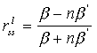
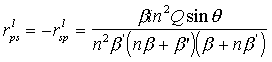
Here, q is the angle of incidence, n is the index of refraction of the film, ![]() , and
, and 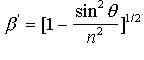 . The term
 ![]() represents the reflection coefficient which relates the incident s-wave to the reflected p-wave in the longitudinal effect. From the transverse coefficients, it is clear that the light does not undergo a rotation of its plane of polarisation, since the off-diagonal terms which give rise to the rotation are equal to zero
represents the reflection coefficient which relates the incident s-wave to the reflected p-wave in the longitudinal effect. From the transverse coefficients, it is clear that the light does not undergo a rotation of its plane of polarisation, since the off-diagonal terms which give rise to the rotation are equal to zero ![]() . The only quantity which is dependent on the magnetisation is the reflection coefficient relating the incident and reflected p-polarised light, as shown/explained by Figure 2.5c, whereas the longitudinal coefficients indicate a rotation of the polarisation by the existence of the off diagonal terms
. The only quantity which is dependent on the magnetisation is the reflection coefficient relating the incident and reflected p-polarised light, as shown/explained by Figure 2.5c, whereas the longitudinal coefficients indicate a rotation of the polarisation by the existence of the off diagonal terms ![]() .
.
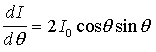
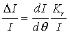
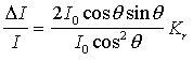
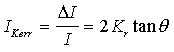
For simplicity and clarity the author has used a pictorial explanation of the Kerr effect for a more qualitative approach here.
| Growth and study of magnetostrictive FeSiBC thin films, for device applications, Mannan Ali (1999) | 8 | |
|---|---|---|
| (Online Copy) |